Your Cart is Empty


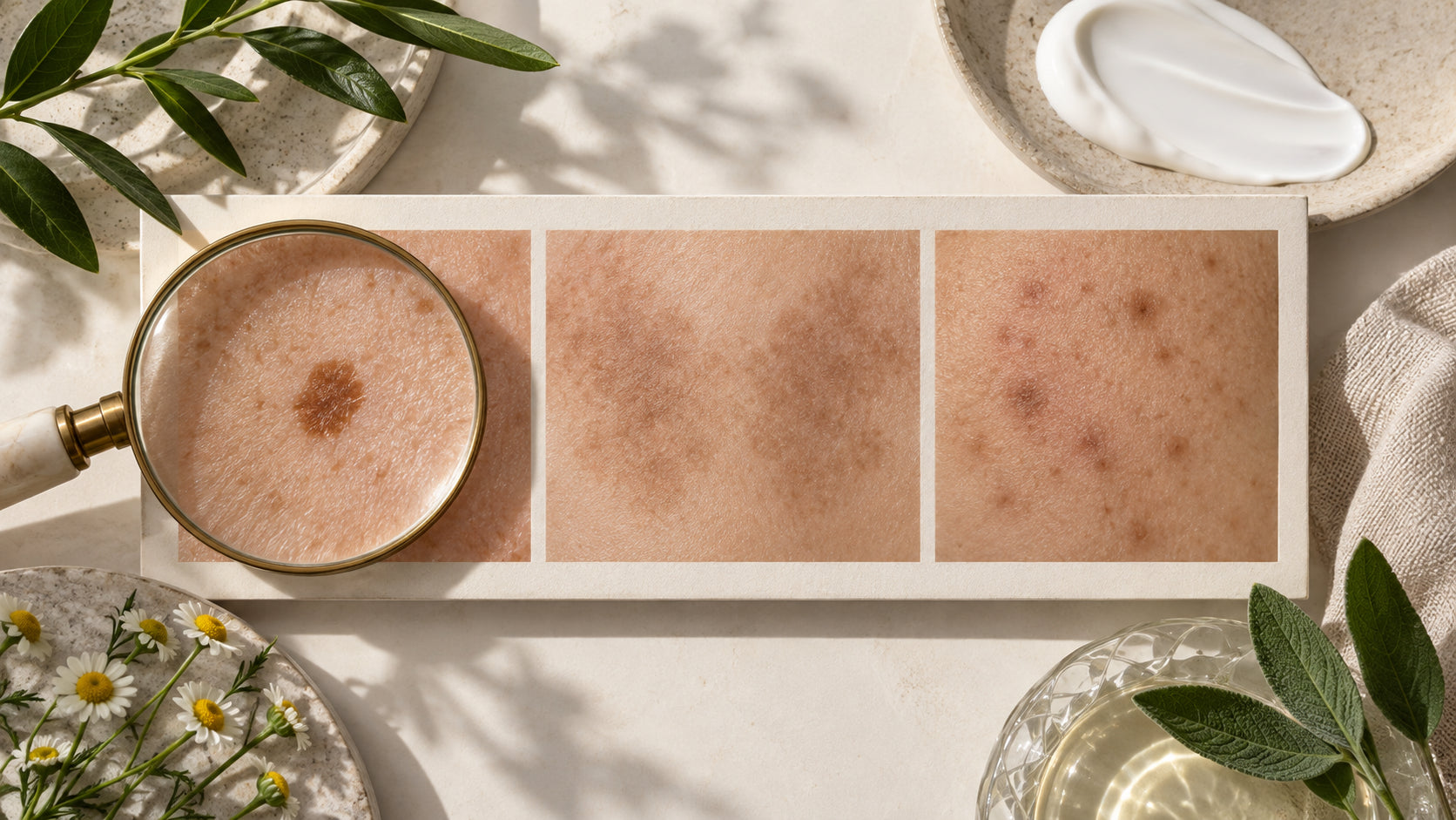
Quick answer: Sun spots are usually localized marks linked with cumulative sun exposure; melasma often forms symmetrical patches; and post-acne marks appear where inflammation healed. These clues are not a diagnosis, and any new or changing lesion should be examined by a clinician.
| Feature | Sun-induced spot | Melasma | Post-acne mark |
|---|---|---|---|
| Pattern | One or more defined marks | Often symmetrical patches | Matches the site of a prior pimple |
| Common areas | Face, hands, chest and other exposed skin | Cheeks, forehead, upper lip and jawline | Any acne-prone area |
| Main context | Cumulative light exposure | Light, hormones and predisposition | Inflammation, picking or acne injury |
| Best first step | Sun protection and assessment if changing | Dermatologist-guided plan | Control acne and avoid picking |
Solar lentigines are benign marks associated with accumulated sun exposure, but skin cancer can also appear as a changing pigmented lesion. The safe approach is assessment of anything new, irregular, evolving, itchy or bleeding—not assuming every “sun spot” is cosmetic.
Melasma is a chronic pigment condition influenced by ultraviolet radiation, visible light, hormones and genetic susceptibility. It can recur and often needs a long-term plan. See our evidence-based melasma guide.
Post-inflammatory hyperpigmentation is flat. A depressed or raised change is a scar and may require professional procedures. Compare them in Acne scars vs. dark spots and follow the post-acne mark routine.
Gentle care, trigger control and daily broad-spectrum sunscreen are foundational. Vitamin C and other brightening ingredients may support appearance, but the appropriate routine depends on the type. See best ingredients for hyperpigmentation.
Only a qualified clinician can diagnose a pigmented lesion. Seek prompt assessment for a spot that changes, bleeds or looks unlike your others.